manual ventilator
Manual Ventilators: A Comprehensive Overview (as of 05/05/2026)
Manual ventilation, a critical skill in emergency medicine, utilizes devices to assist or replace spontaneous breathing, offering a lifeline when natural respiration fails.

Today’s date is 05/05/2026 02:00:14, reflecting the current relevance of understanding these vital techniques and technologies.
Manual ventilation represents a cornerstone of emergency respiratory support, bridging the gap when a patient’s own breathing is inadequate or ceases altogether. This technique, employing a manual ventilator – often a bag-valve-mask (BVM) – delivers breaths directly to the patient’s lungs, ensuring oxygenation and ventilation.
It’s a fundamental skill for healthcare professionals, requiring proficiency in proper technique to avoid complications. Unlike mechanical ventilation, manual methods rely entirely on the operator’s actions, demanding constant vigilance and adaptation to the patient’s needs.
Understanding the principles of manual ventilation is crucial in diverse settings, from pre-hospital emergency care to operating rooms and intensive care units. Effective implementation can be life-saving, providing critical time until definitive airway management or spontaneous breathing resumes.
What is a Manual Ventilator?
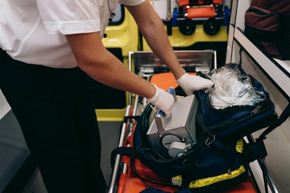
A manual ventilator, commonly known as a bag-valve-mask (BVM) or Ambu bag, is a handheld device used to provide positive pressure ventilation to patients. It consists of a reservoir bag, a valve system, and a mask that creates a seal over the patient’s mouth and nose, or connects to an endotracheal tube.
The device functions by the operator squeezing the bag, delivering a pre-defined volume of air into the patient’s lungs. The one-way valve prevents exhaled air from re-entering the bag, ensuring fresh gas delivery.
Unlike automated systems, a manual ventilator requires direct human operation, offering immediate control but demanding continuous effort and skill. It’s a portable, reliable tool for emergency situations.
Historical Development of Manual Ventilation
The origins of manual ventilation trace back to the late 19th century, with early attempts focusing on mouth-to-mouth resuscitation and mechanical devices to assist breathing. Significant advancements occurred during the polio epidemics of the mid-20th century, driving the need for reliable ventilation support.
The development of the bag-valve-mask (BVM) in the 1950s, attributed to Dr; Heinrich Bouman and others, revolutionized emergency respiratory care. This portable, relatively simple device offered a significant improvement over previous methods.
Over the decades, refinements in mask design, bag materials, and valve technology have enhanced the effectiveness and safety of manual ventilation, solidifying its role in modern medicine.
Key Components of a Manual Ventilator
A typical manual ventilator, often a bag-valve-mask (BVM) system, comprises several essential components. The bellows, or bag, delivers pressurized air to the patient. A one-way valve prevents exhaled air from re-entering the bag. The mask or endotracheal tube establishes an airtight seal with the patient’s airway.
Crucially, a pressure gauge monitors the force of air delivered, while a flow meter (sometimes integrated) indicates the volume of air per breath. An oxygen reservoir attached to the system allows for supplemental oxygen delivery, enriching the air mixture.
These components work in concert to provide controlled, assisted ventilation.
Bellows and Reservoir Bag
The bellows, the core of a manual ventilator, is a self-inflating bag that healthcare professionals compress to deliver breaths. Constructed from durable materials like latex or silicone, it stores a pre-determined volume of air. Connected to the bellows is the reservoir bag, a crucial component for supplemental oxygenation.
This reservoir acts as a holding chamber, ensuring a high concentration of oxygen is available for each breath delivered. Properly filling the reservoir before each squeeze is vital for maintaining adequate oxygen levels. The size of the reservoir bag is selected based on the patient’s size and ventilatory needs.
Airway Connection & Mask/Endotracheal Tube
Establishing a secure airway connection is paramount during manual ventilation. This is achieved using either a mask or an endotracheal tube (ETT). Masks, available in various sizes, create a seal over the patient’s mouth and nose, suitable for short-term ventilation.
An ETT, inserted into the trachea, provides a definitive airway, ideal for prolonged ventilation. Proper sizing and insertion of the ETT are critical to prevent complications. The connection between the ventilator and the airway utilizes a standardized adapter, ensuring a leak-proof seal. Regular assessment of the airway is essential to maintain effective ventilation.
Pressure Gauge & Flow Meter
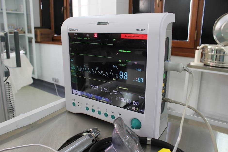
The pressure gauge on a manual ventilator is crucial for monitoring the force delivered with each breath, preventing barotrauma. It displays airway pressure in centimeters of water (cmH2O), guiding clinicians to avoid excessive inflation. Simultaneously, the flow meter indicates the rate at which gas is delivered, typically measured in liters per minute (L/min).
These instruments allow for precise control and adjustment of ventilation parameters. Observing both readings helps optimize tidal volume and ensures adequate oxygenation. Regular monitoring of these values is vital for patient safety and effective respiratory support during manual ventilation.
Principles of Operation: How Manual Ventilation Works
Manual ventilation operates on the principle of creating positive pressure within the patient’s airway. The user compresses a bellows, forcing air from a reservoir into the lungs. This overcomes the patient’s inability to breathe adequately on their own.
The process mimics natural breathing by delivering a controlled tidal volume – the amount of air inhaled or exhaled with each breath. Successful ventilation requires a tight airway seal and coordinated compression and release of the bellows, ensuring oxygen delivery and carbon dioxide removal. Proper technique is paramount for effective gas exchange.
Positive Pressure Ventilation Explained
Positive Pressure Ventilation (PPV), central to manual ventilation, involves delivering breaths into the patient’s lungs using external pressure. Unlike natural breathing, which relies on negative pressure created by the diaphragm, PPV actively pushes air into the airways.

This is achieved by manually compressing a bag-valve-mask or utilizing a mechanical ventilator. PPV effectively bypasses airway obstruction or muscle weakness, ensuring oxygenation and ventilation. Careful monitoring of tidal volume and airway pressure is crucial to avoid lung injury, making controlled delivery essential for optimal patient outcomes.
Understanding Tidal Volume and Respiratory Rate
Tidal Volume (TV) represents the volume of air delivered with each breath during manual ventilation, typically ranging from 6-8 ml/kg of ideal body weight. Respiratory Rate (RR) defines the number of breaths delivered per minute, usually between 10-12 for adults.
These two parameters are fundamental to effective ventilation. Adjusting TV and RR allows clinicians to regulate carbon dioxide levels (EtCO2) and oxygenation. Insufficient TV leads to hypoventilation, while excessive TV risks volutrauma. Proper synchronization with the patient’s intrinsic rhythm optimizes ventilation and minimizes complications, ensuring adequate gas exchange.
Indications for Manual Ventilation
Manual ventilation becomes crucial in scenarios where a patient cannot maintain adequate breathing independently. Respiratory failure, stemming from conditions like pneumonia or COPD, is a primary indication. Trauma patients with chest injuries or altered mental status often require assisted ventilation.

Post-operative care frequently necessitates temporary support, particularly after procedures affecting respiratory function. Emergency situations, including drug overdose or airway obstruction, demand immediate intervention. Recognizing these indications and initiating prompt manual ventilation can be life-saving, bridging the gap until definitive airway management is established.
Respiratory Failure – Types and Causes
Respiratory failure manifests in diverse forms, demanding tailored ventilation strategies. Hypoxemic failure arises from insufficient oxygen transfer, often due to pneumonia or pulmonary edema. Hypercapnic failure results from inadequate carbon dioxide removal, linked to COPD or neuromuscular diseases.
Underlying causes are varied, encompassing infections, chronic conditions, and acute events like asthma exacerbations. Trauma, airway obstruction, and drug overdose can also precipitate respiratory failure. Accurate identification of the failure type and its root cause is paramount for effective manual ventilation and subsequent treatment.
Trauma and Emergency Situations
Trauma scenarios, such as chest injuries or severe head trauma, frequently necessitate immediate manual ventilation. Emergency situations like drowning, electrocution, and opioid overdose often compromise respiratory function, requiring prompt intervention.
Manual ventilation bridges the gap until definitive airway management can be established. It provides crucial oxygenation and ventilation, preventing further deterioration. Rapid assessment of the patient’s condition and skillful application of manual ventilation techniques are vital in these critical moments, potentially saving lives while awaiting advanced medical support.
Post-Operative Respiratory Support
Post-operative respiratory support often involves manual ventilation, particularly after surgeries affecting the chest, abdomen, or those requiring prolonged anesthesia. Residual effects of anesthetic drugs can suppress spontaneous breathing, demanding temporary ventilatory assistance.
Manual ventilation serves as a crucial transitional phase, allowing patients to gradually regain respiratory control. It’s frequently employed during emergence from anesthesia and in the immediate post-operative period, ensuring adequate oxygenation and preventing hypoventilation. Careful monitoring and adjustment of ventilation parameters are essential for a smooth recovery.
Techniques of Manual Ventilation
Effective manual ventilation relies on mastering specific techniques. Proper hand placement on the bellows is paramount, utilizing a two-handed grip for consistent, controlled compressions. A rhythmic squeeze, avoiding excessive force, delivers optimal tidal volume.
Synchronizing ventilation with the patient’s intrinsic breathing efforts is crucial, minimizing resistance and potential trauma. Observing chest rise and fall provides visual feedback on ventilation effectiveness. Adjusting ventilation rate and tidal volume based on patient response and clinical assessment is vital for maintaining appropriate gas exchange.
Proper Hand Placement and Bellows Squeeze
Correct hand positioning is fundamental to effective manual ventilation. A ‘C-E’ grip – encircling the bellows with the thumb and index finger forming a ‘C’ and the remaining fingers forming an ‘E’ – provides optimal control.
The bellows squeeze should be slow and deliberate, avoiding rapid compressions which can cause gastric distension. Aim for a smooth, consistent delivery of air, observing chest rise as a guide. Avoid excessive force; the goal is adequate ventilation, not forceful inflation. Regular reassessment of technique ensures continued effectiveness.
Synchronizing Ventilation with Patient Breathing
Effective manual ventilation often requires synchronizing breaths with the patient’s own respiratory efforts. This minimizes resistance and reduces the risk of complications. Observe for signs of spontaneous breathing – chest rise, abdominal movement – and deliver breaths with these efforts, not against them.
Asynchronous ventilation can lead to patient discomfort and increased airway pressure. Gentle breaths, coupled with attentive observation, facilitate synchronization. If the patient is fighting the ventilator, consider adjusting the rate or depth of breaths, or providing sedation if appropriate and within your scope of practice.
Ventilation Rate and Tidal Volume Adjustment
Adjusting ventilation rate and tidal volume is crucial for optimal oxygenation and ventilation. A typical starting point is a rate of 10-12 breaths per minute, with a tidal volume of 6-8 mL/kg of ideal body weight.

Regularly assess the patient’s clinical status – including pulse oximetry, respiratory effort, and arterial blood gases (if available) – to guide adjustments. Increased respiratory rate may be needed for increased metabolic demand, while tidal volume adjustments impact CO2 elimination. Careful titration is key to avoid over or under-ventilation.

Potential Complications of Manual Ventilation
Manual ventilation, while life-saving, carries potential risks. Barotrauma, lung injury from excessive pressure, and volutrauma, damage from large tidal volumes, are significant concerns.
Gastric distension, caused by air entering the stomach, can compromise diaphragm function and increase aspiration risk. Vigilant monitoring for signs of hypoxia (low oxygen) or hyperoxia (high oxygen) is essential, as both can be detrimental. Proper technique and careful pressure control minimize these complications, emphasizing the need for skilled operators.
Barotrauma and Volutrauma
Barotrauma, lung injury resulting from excessive airway pressure during manual ventilation, can manifest as pneumothorax or subcutaneous emphysema. Careful pressure monitoring and adherence to recommended limits are crucial preventative measures.
Volutrauma, conversely, stems from overdistension of the alveoli due to large tidal volumes. This causes inflammatory damage and can lead to acute respiratory distress syndrome (ARDS). Utilizing appropriate tidal volume settings, guided by patient characteristics, minimizes this risk. Recognizing the delicate balance between oxygenation and lung protection is paramount.
Gastric Distension
Gastric distension is a common complication during manual ventilation, particularly with positive pressure. Air entering the stomach can cause discomfort, impede diaphragm movement, and increase the risk of aspiration.
Strategies to mitigate this include ensuring proper endotracheal tube placement, utilizing appropriate ventilation rates and tidal volumes, and considering the use of a cricoid pressure maneuver. Frequent auscultation of the stomach can help detect early signs of distension. Prompt decompression via a nasogastric tube may be necessary to alleviate symptoms and prevent further complications, safeguarding patient comfort and respiratory function.
Hypoxia and Hyperoxia

Hypoxia, insufficient oxygen, and hyperoxia, excessive oxygen, both pose risks during manual ventilation. Inadequate ventilation or low inspired oxygen concentrations can lead to hypoxia, causing tissue damage. Conversely, prolonged exposure to high oxygen levels can generate reactive oxygen species, contributing to lung injury.
Careful monitoring of oxygen saturation via pulse oximetry is crucial. Adjusting the inspired oxygen concentration (FiO2) based on patient needs and arterial blood gas analysis is essential. Aiming for normoxia – maintaining adequate oxygen levels without excess – optimizes patient outcomes and minimizes potential harm from either extreme.
Manual Ventilation vs. Mechanical Ventilation: A Comparison
Manual ventilation offers immediate respiratory support, requiring constant operator effort and skill, while mechanical ventilation delivers breaths automatically via a machine. Manual methods are ideal for short-term stabilization or when mechanical ventilation is unavailable.
Mechanical ventilation provides consistent, adjustable parameters, reducing operator fatigue and allowing for precise control of ventilation. However, it necessitates specialized equipment and monitoring. Manual ventilation is portable and doesn’t rely on power, making it valuable in resource-limited settings or transport. Both methods aim to improve oxygenation and ventilation, but differ in complexity and reliance on technology.
Advantages of Manual Ventilation
Manual ventilation provides a crucial, readily available method for respiratory support, especially in emergencies where immediate intervention is vital. Its portability and lack of reliance on electricity make it invaluable during transport or in locations lacking power sources.
Furthermore, it offers a tactile connection with the patient, allowing clinicians to assess lung compliance and response to ventilation. This direct feedback is absent in automated systems. Manual ventilation is also cost-effective, requiring minimal equipment compared to mechanical ventilators. It’s a fundamental skill, enhancing a clinician’s ability to manage respiratory distress effectively.
Disadvantages of Manual Ventilation
Manual ventilation demands significant physical exertion from the operator, leading to fatigue, particularly during prolonged resuscitation efforts. This fatigue can compromise ventilation quality and consistency. Maintaining precise tidal volumes and respiratory rates relies heavily on the operator’s skill and experience, introducing potential for error.
Unlike mechanical ventilation, manual methods lack automated alarms for pressure or volume limits, increasing the risk of barotrauma or volutrauma. It also requires continuous, dedicated personnel, which can be a limitation in resource-constrained environments. The absence of data logging hinders detailed respiratory assessment.
Training and Certification for Manual Ventilation
Comprehensive training is paramount for effective manual ventilation. Initial courses, like Basic Life Support (BLS) and Advanced Cardiac Life Support (ACLS), introduce fundamental skills, but dedicated respiratory training is crucial. These programs emphasize proper technique, airway management, and recognizing potential complications.
Simulation-based training, utilizing realistic manikins, allows healthcare professionals to practice in controlled environments, refining their skills and building confidence. Certification programs, often offered by professional organizations, validate competency. Regular refresher courses are vital to maintain proficiency and stay updated on best practices, ensuring optimal patient care.
Future Trends in Manual Ventilation Technology
Innovations in materials are leading to lighter, more durable manual ventilators, enhancing portability and ease of use in challenging environments. Integration of smart technology, including sensors for real-time feedback on tidal volume and pressure, is gaining traction. These devices will offer improved accuracy and reduce operator fatigue.
Research focuses on developing ventilators with automated assistance features, adapting to patient needs dynamically. Furthermore, advancements in mask design aim to improve seal and reduce leaks, optimizing ventilation effectiveness. Telemedicine applications, enabling remote guidance during manual ventilation, represent a promising future direction.